Services
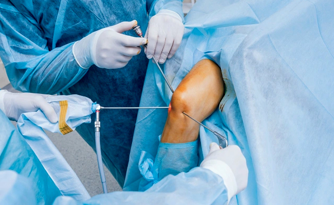
ARTHROSCOPY
Arthroscopy is a surgical procedure doctors use to look at, diagnose, and treat problems
inside a joint. Doctor may recommend it if you have inflammation in a joint, have injured a
joint, or have damaged a joint over time. You can have arthroscopy on any joint. Most often,
it’s done on the knee, shoulder, elbow, ankle, hip, or wrist.It is a procedure which is done through minimul incision with the minor instrument without
opeming the joint. It is a keyhole Surgery.
During the procedure, the doctor will insert a tool called an arthroscope into your joint
through several small cuts to see how much damage is in the joint. They can also repair many
injuries during arthroscopy.
WHAT HAPPENS DURING THE PROCEDURE ?
Your doctor will perform arthroscopic surgery in a hospital or outpatient operating room. The
type of anesthesia you’ll receive depends on the joint and what your surgeon suspects is the
problem. It may be general anesthesia (you’ll be asleep during surgery), or your doctor will
give it to you through your spine. They might also numb the area they are doing the surgery
on.
Your doctor will insert special pencil-thin instruments through a small cut (incision) the
size of a buttonhole. The arthroscope tool they use has a camera lens and a light. It allows
them to see inside the joint. The camera projects an image of the joint onto a screen. The
surgeon will fill the joint with sterile fluid to widen it so it’s easier to see.
They’ll look inside the joint, diagnose the problem, and decide what type of surgery you
need, if any. If you do need surgery, your surgeon will insert special tools through other
small incisions called portals. They’ll use them to cut, shave, grasp, and anchor stitches
into bone.
If your surgeon decides you need traditional, “open” surgery to fix the problem, they may do
it at the same time as your arthroscopic surgery.
Afterward, they’ll remove the arthroscope and any attachments. They’ll close the wound with
special tape or stitches.
WHAT ABOUT RECOVERY ?
Arthroscopic surgery usually results in less joint pain and stiffness than open surgery.
Recovery also generally takes less time. When the arthroscopy is over, you'll be taken to a
recovery room where you'll rest for about an hour or more. You may have some pain in the
joint after surgery. Your doctor may prescribe pain medication and exercise. They might also
prescribe aspirin or other medication to prevent blood clots.
Apply ice for the first 24 hours to reduce swelling. If you've had arthroscopy on your knee,
elevate the leg to reduce pain. Take pain medicines as prescribed, and do not drink alcohol.
You may need crutches, a splint, or a sling for support as you recover. Rehabilitation or
specific exercises can help speed your recovery. Your doctor will tell you which ones are
safe to do.
ACL
ACL refers to the anterior cruciate ligament, which is one of the major ligaments in the
knee. ACL injuries often occur during activities involving sudden stops, changes in
direction, or direct blows to the knee. These injuries are commonly seen in sports such as
soccer, basketball, and skiing.
Arthroscopic ACL reconstruction is a surgical procedure performed to repair a torn ACL.
During the surgery, the surgeon removes the damaged ligament and replaces it with a graft,
typically taken from another part of the patient's body or from a donor. The arthroscope
helps the surgeon visualize and navigate the knee joint during the procedure, resulting in
smaller incisions, reduced tissue damage, and potentially faster recovery compared to
traditional open surgery.
PCL
PCL arthroscopy, also known as posterior cruciate ligament arthroscopy, is a surgical
procedure performed to diagnose and treat injuries or conditions affecting the posterior
cruciate ligament (PCL) in the knee joint. The PCL is one of the four major ligaments in the
knee and plays a crucial role in stabilizing the joint.
PCL arthroscopy is a minimally invasive technique compared to traditional open surgery. It
offers several potential benefits, such as smaller incisions, reduced pain, faster recovery,
and shorter hospital stays. However, the suitability of arthroscopy depends on the specific
characteristics of the PCL injury and the patient's individual circumstances.
Meniscus Arthroscopy Enjury
A meniscus arthroscopy injury refers to a condition where the meniscus, which is a C-shaped
cartilage structure in the knee joint, is damaged and requires arthroscopic surgery for
treatment. Arthroscopy is a minimally invasive surgical procedure that involves the use of a
tiny camera, called an arthroscope, to visualize and treat problems inside a joint.
In the case of a meniscus arthroscopy injury, the meniscus may be torn or damaged due to
various reasons, such as sports-related activities, trauma, or degenerative changes in the
knee joint. The meniscus plays a crucial role in cushioning and stabilizing the knee joint,
and when injured, it can cause pain, swelling, stiffness, and difficulty in moving the knee.
Shoulder Rotator cuff care
A shoulder rotator cuff arthroscopy refers to a surgical procedure performed to diagnose and
treat problems with the rotator cuff in the shoulder joint using an arthroscope. The rotator
cuff is a group of four muscles and tendons that surround the shoulder joint, providing
stability and facilitating movement.
When the rotator cuff is injured or damaged, it can lead to pain, weakness, and limited
range of motion in the shoulder. Common causes of rotator cuff injuries include repetitive
overhead activities, trauma, and age-related degeneration.
Bankart
Bankart: Bankart refers to a specific type of shoulder injury known as a Bankart lesion or
Bankart tear. It involves the detachment of a piece of cartilage called the labrum from the
front of the shoulder joint. This injury often occurs in association with shoulder
dislocations or instability. Bankart repairs are commonly performed arthroscopically to
reattach the labrum to the shoulder socket.
Hill-Sachs
Hill-Sachs: Hill-Sachs lesion is another type of shoulder injury that often occurs in
conjunction with a Bankart tear. It is a compression fracture or indentation on the back of
the upper arm bone (humerus) caused by contact with the edge of the shoulder socket during a
dislocation. Hill-Sachs lesions can contribute to shoulder instability and may require
treatment during a Bankart repair surgery.